博文
科学家解决了几十年的显微镜问题  精选
精选
||
科学家解决了几十年的显微镜问题
诸平
据荷兰代尔夫特理工大学(Delft University Of Technology, Delft, The Netherlands)2024年4月22日提供的消息,科学家解决了几十年的显微镜问题(Scientists Solve Decades-Old Microscopy Problem)。
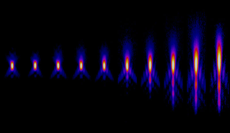
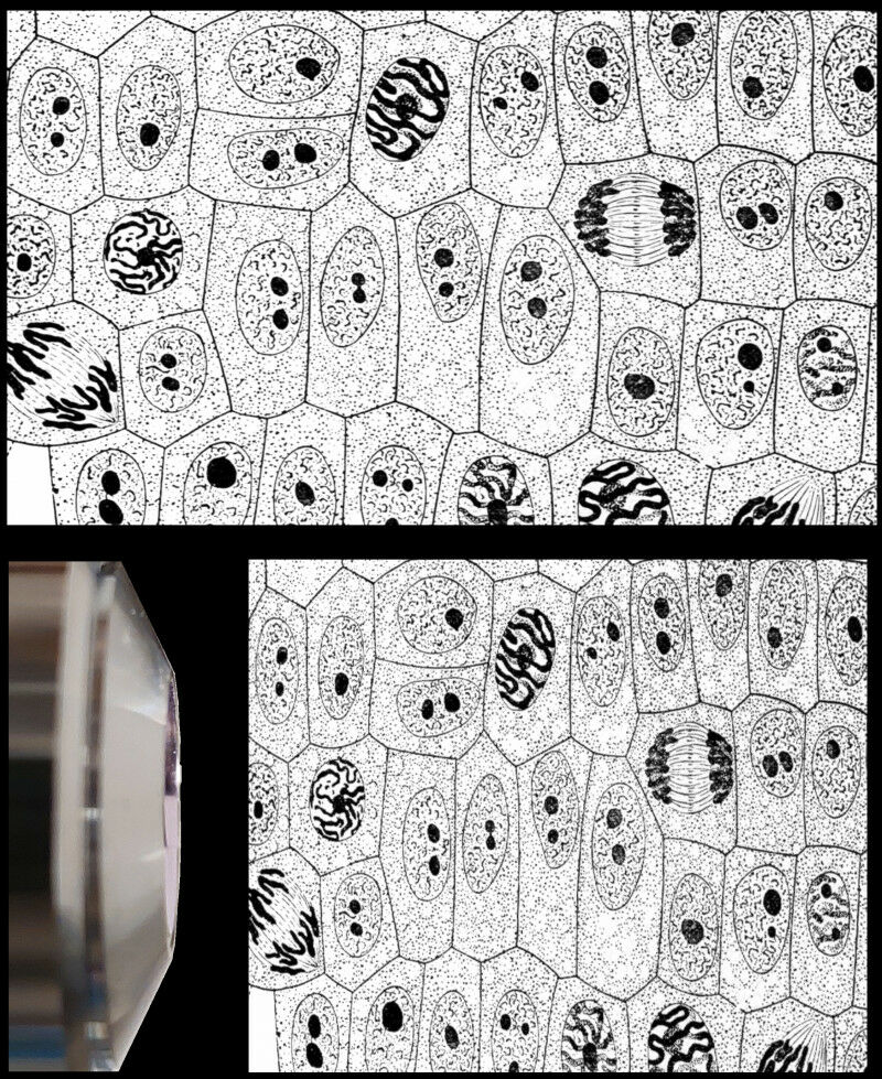
在显微镜下研究组织、细胞和蛋白质对于疾病的预防和治疗至关重要。这项研究需要精确测量这些生物结构的尺寸。然而,当通过光学显微镜(light microscope)观察时,这些样品有时会显得比它们的真实形状更平坦。
代尔夫特理工大学(Delft University of Technology)的研究人员现在首次证明,这种扭曲不是恒定的,这与许多科学家几十年来的假设相反。2024年4月19日发表在《光学》(Optica)杂志网站的这一突破,证实了诺贝尔奖(The Nobel Prize in Chemistry 2014)得主斯蒂芬·赫尔(Nobel laureate Stefan W. Hell)在20世纪90年代的预测。有了在线计算工具和软件,每个研究人员现在都可以确定生物样本的正确深度。原文详见:S. V. Loginov, D. B. Boltje, M. N. F. Hensgens, J. P. Hoogenboom, E. B. van der Wee. Depth-dependent scaling of axial distances in light microscopy. Optica, 2024, 11(4): 553-568. DOI: doi:10.1364/OPTICA.520595. Published 19 April 2024. https://doi.org/10.1364/OPTICA.520595
参与此项研究的除了来自荷兰代尔夫特理工大学的研究人员之外,还有来自荷兰代尔夫特Delmic B.V.公司(Delmic B.V., Delft, The Netherlands)以及荷兰乌得勒支大学(Utrecht University, Utrecht, The Netherlands)的研究人员。
扁平的样本(Flattened sample)
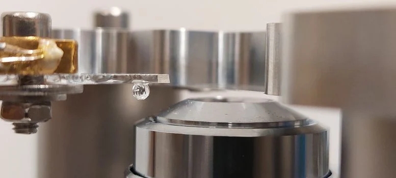
当用显微镜观察生物样品时,如果物镜的透镜处于与样品不同的介质中,光束就会受到干扰。例如,当用被空气包围的透镜观察含水样品时,光线在透镜周围的空气中比在水中弯曲得更剧烈。这种扰动导致样品的测量深度小于实际深度。
因此,样品看起来是扁平的。雅各布·胡根布姆(Jacob Hoogenboom) 副教授解释说:“这个问题已经存在很长时间了,自20世纪80年代以来,人们已经发展了一些理论来确定测定深度的校正系数。然而,所有这些理论都假设这个因素是恒定的,无论样本的深度如何。尽管后来的诺贝尔奖得主斯蒂芬·赫尔在20世纪90年代指出,这种缩放可能与深度有关,但这种情况还是发生了。”
计算,实验和网络工具(Calculations, experiments, and web tool)
前代尔夫特理工大学博士后谢尔盖·洛吉诺夫(Sergey Loginov)通过计算和数学模型证明,样品在靠近透镜的地方确实比远离透镜的地方更平坦。博士候选人达恩·博尔杰(Daan Boltje)和博士后欧内斯特·范德威(Ernest van der Wee,现在荷兰乌得勒支大学工作)随后在实验室中证实,校正因子与深度有关。
欧内斯特·范德威说:“我们已经将我们的研究结果汇编成一个网络工具和软件,随文章一起提供。有了这些工具,任何人都可以为他们的实验确定精确的校正系数。”
了解异常和疾病(Understanding abnormalities and diseases)
“部分得益于我们的计算工具,我们现在可以非常精确地从生物系统中切割出蛋白质及其周围环境,并用电子显微镜确定其结构。这种显微镜非常复杂,耗时,而且非常昂贵。因此,确保你看到的是正确的结构非常重要,”达恩·博尔杰说。
“有了更精确的深度测定,我们需要花更少的时间和金钱在那些没有达到生物目标的样本上。最终,我们可以研究更多相关的蛋白质和生物结构。确定生物系统中蛋白质的精确结构对于理解并最终对抗异常和疾病至关重要。”
关于网络工具(About the web tool)
在网络工具中(In the web tool),您可以填写实验的相关细节,例如折射率(refractive indices)、物镜的孔径角(aperture angle)和所用光的波长(wavelength)。然后,该工具显示与深度相关的缩放因子(scaling factor)曲线。您还可以导出这些数据以供自己使用。此外,您可以将结果与每个现有理论的结果结合起来绘图。
这项工作是Cryo3Beams项目(Cryo3Beams project)的一部分,该项目由荷兰研究委员会(Dutch Research Council / NWO)应用与工程科学领域的高科技系统和材料计划{ High Tech Systems and Materials programme of the Applied and Engineering Sciences domain of the Dutch Research Council (NWO)}资助。.
上述介绍,仅供参考。欲了解更多信息,敬请注意浏览原文或者相关报道。
2014年诺贝尔化学奖得主Stefan Hell的代表性论文是一篇睡美人文献
In volume fluorescence microscopy, refractive index matching is essential to minimize aberrations. There are, however, common imaging scenarios where a refractive index mismatch (RIM) between immersion and a sample medium cannot be avoided. This RIM leads to an axial deformation in the acquired image data. Over the years, different axial scaling factors have been proposed to correct for this deformation. While some reports have suggested a depth-dependent axial deformation, so far none of the scaling theories has accounted for a depth-dependent, non-linear scaling. Here, we derive an analytical theory based on determining the leading constructive interference band in the objective lens pupil under RIM. We then use this to calculate a depth-dependent re-scaling factor as a function of the numerical aperture (NA), the refractive indices n1 and n2, and the wavelength λ. We compare our theoretical results with wave-optics calculations and experimental results obtained using a measurement scheme for different values of NA and RIM. As a benchmark, we recorded multiple datasets in different RIM conditions, and corrected these using our depth-dependent axial scaling theory. Finally, we present an online web applet that visualizes the depth-dependent axial re-scaling for specific optical setups. In addition, we provide software that will help microscopists to correctly re-scale the axial dimension in their imaging data when working under RIM.
https://blog.sciencenet.cn/blog-212210-1430856.html
上一篇:前所未有的光波:科学家揭开了突破性的光量子探测
下一篇:最大的癌症全基因组测序研究


