博文
更好地了解癌症和心脏病
||
更好地了解癌症和心脏病
诸平
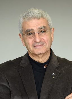
Fig. 2 Nabil G. Seidah, CREDIT: IRCM
据加拿大蒙特利尔大学(Université de Montréal)2023年1月19日报道,更好地了解癌症和心脏病( Better understanding cancer and heart disease)。
一个由加拿大领导的研究小组最终确定了一种关键蛋白质调节低密度脂蛋白胆固醇(LDL cholesterol)的分子机制。在了解心血管疾病和某些癌症相关机制的关键一步中,加拿大领导的研究团队取得了世界第一的成功:他们发现了蛋白质PCSK9降解低密度脂蛋白受体,血液中最丰富的胆固醇颗粒。
蒙特利尔临床研究所(Institut de recherches cliniques de Montréal简称IRCM / Montreal Clinical Research Institute)生化神经内分泌学研究室(biochemical neuroendocrinology research unit)主任、蒙特利尔大学医学教授纳比尔·赛达(Nabil G. Seidah)的发现,于2022年12月22日已经在《分子代谢》(Molecular Metabolism)杂志网站发表——Carole Fruchart Gaillard, Ali Ben Djoudi Ouadda, Lidia Ciccone, Emmanuelle Girard, Sepideh Mikaeeli, Alexandra Evagelidis, Maïlys Le Dévéhat, Delia Susan-Resiga, Evelyne Cassar Lajeunesse, Hervé Nozach, Oscar Henrique Pereira Ramos, Aurélien Thureau, Pierre Legrand, Annik Prat, Vincent Dive, Nabil G. Seidah. Molecular interactions of PCSK9 with an inhibitory nanobody, CAP1 and HLA-C: Functional regulation of LDLR levels. Molecular Metabolism, January 2023, Volume 67, 101662. DOI: 10.1016/j.molmet.2022.101662. Epub 2022 Dec 22. https://doi.org/10.1016/j.molmet.2022.101662
参与此项研究的有来自法国巴黎萨克雷大学(Université Paris-Saclay, France)、法国SOLEIL同步加速器(Synchrotron SOLEIL, Saint-Aubin, France);加拿大蒙特利尔大学蒙特利尔临床研究所(Montreal Clinical Research Institute, affiliated to the University of Montreal, Quebec, Canada)以及意大利比萨大学(University of Pisa, Via Bonanno, Italy)的研究人员。
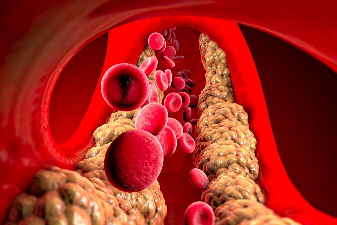
低密度脂蛋白(Low density lipoproteins简称LDL)会在血液中积聚并导致动脉粥样硬化和心脏病。LDL和与之相关的胆固醇 (LDLc) 的水平直接受LDL受体(LDL receptors简称LDLR) 从血流中收集LDL并将其内化(主要进入肝脏细胞)的能力调节。表面LDLR将LDL驱动到它被捕获的细胞中,然后LDLR返回表面进行另一轮捕获。
与PCSK9蛋白相关的罕见病例(Rare cases linked to the PCSK9 protein)
大多数家族性高胆固醇血症病例与LDLR功能障碍有关。但罕见的病例与纳比尔·赛达实验室于2003年发现的PCSK9蛋白质(PCSK9 protein)有关。PCSK9也存在于血液中,它与 LDLR结合并促进其被肝细胞降解,阻止其返回表面捕获LDL。一些高胆固醇血症患者有一个“超级PCSK9(super PCSK9)”,可以增强LDLR的降解。
近年来,患者可以使用高效的治疗方法抑制 PCSK9 的功能(称为单克隆抗体)或降低血流中PCSK9的水平(称为RNAi),从而产生更大量的LDLR,确保LDLc降低与传统他汀类药物相比减少超过60%。
现在,纳比尔·赛达和他的团队的工作揭开了之前被误解的机制的面纱,即PCSK9将LDLR 拖向溶酶体(lysosomes),细胞在溶酶体中降解PCSK9-LDLR复合物。
三种伙伴蛋白的复合体(A complex of three partner proteins)
在他们的实验室中,纳比尔·赛达和他的团队进行了结构分析,揭示了3种 PCSK9伙伴蛋白(PCSK9 partner proteins)复合物的形成,包括LDLR、CAP1和HLA-C。
作为免疫系统中的关键蛋白质,HLA-C被发现发挥着关键作用:它将整个复合体引导至溶酶体。HLA-C可以识别“自我”,也可以刺激T淋巴细胞(T lymphocytes)的抗肿瘤活性。
就其本身而言,PCSK9通过增加细胞表面的HLA-C水平来帮助防止肿瘤的生长和相关的转移。最终,希望能够开发出抑制剂来阻止PCSK9和HLA-C的相互作用,并阻断PCSK9在LDLR和HLA-C上的功能。这一突破随后可以应用于临床实践,以治疗心血管疾病以及各种类型的癌症和转移患者。
本研究得到了CIHR 基金会(CIHR Foundation, NGS: #148363)、加拿大前体蛋白水解主席(Canada Chair in Precursor Proteolysis, NGS: #950-231335)和 Leducq 基金会(Fondation Leducq, NGS: #13 CVD 03)的资助。
上述介绍,仅供参考。欲了解更多信息,敬请注意浏览原文或者相关报道。
• Structure of PCSK9/CHRD and an inhibitory nanobody that prevents LDLR degradation.
• Binding of CAP1 to PCSK9 enhances its activity and abrogates nanobody inhibition.
• CAP1-binding exposes PCSK9's M2 domain thereby enhancing PCSK9-LDLR degradation.
• CAP1-PCSK9 recruits an MHC-I-like protein to sort CAP1-PCSK9-LDLR to lysosomes.
• HLA-C is likely a “protein X” critical for PCSK9 functional degradation of the LDLR.
Objective
The liver-derived circulating PCSK9 enhances the degradation of the LDL receptor (LDLR) in endosomes/lysosomes. PCSK9 inhibition or silencing is presently used in clinics worldwide to reduce LDL-cholesterol, resulting in lower incidence of cardiovascular disease and possibly cancer/metastasis. The mechanism by which the PCSK9-LDLR complex is sorted to degradation compartments is not fully understood. We previously suggested that out of the three M1, M2 and M3 subdomains of the C-terminal Cys/His-rich-domain (CHRD) of PCSK9, only M2 is critical for the activity of extracellular of PCSK9 on cell surface LDLR. This likely implicates the binding of M2 to an unknown membrane-associated “protein X” that would escort the complex to endosomes/lysosomes for degradation. We reported that a nanobody P1.40 binds the M1 and M3 domains of the CHRD and inhibits the function of PCSK9. It was also reported that the cytosolic adenylyl cyclase-associated protein 1 (CAP1) could bind M1 and M3 subdomains and enhance the activity of PCSK9. In this study, we determined the 3-dimensional structure of the CHRD-P1.40 complex to understand the intricate interplay between P1.40, CAP1 and PCSK9 and how they regulate LDLR degradation.
Methods
X-ray diffraction of the CHRD-P1.40 complex was analyzed with a 2.2 Å resolution. The affinity and interaction of PCSK9 or CHRD with P1.40 or CAP1 was analyzed by atomic modeling, site-directed mutagenesis, bio-layer interferometry, expression in hepatic cell lines and immunocytochemistry to monitor LDLR degradation. The CHRD-P1.40 interaction was further analyzed by deep mutational scanning and binding assays to validate the role of predicted critical residues. Conformational changes and atomic models were obtained by small angle X-ray scattering (SAXS).
Results
We demonstrate that PCSK9 exists in a closed or open conformation and that P1.40 favors the latter by binding key residues in the M1 and M3 subdomains of the CHRD. Our data show that CAP1 is well secreted by hepatic cells and binds extracellular PCSK9 at distinct residues in the M1 and M3 modules and in the acidic prodomain. CAP1 stabilizes the closed conformation of PCSK9 and prevents P1.40 binding. However, CAP1 siRNA only partially inhibited PCSK9 activity on the LDLR. By modeling the previously reported interaction between M2 and an R-X-E motif in HLA-C, we identified Glu567 and Arg549 as critical M2 residues binding HLA-C. Amazingly, these two residues are also required for the PCSK9-induced LDLR degradation.
Conclusions
The present study reveals that CAP1 enhances the function of PCSK9, likely by twisting the protein into a closed configuration that exposes the M2 subdomain needed for targeting the PCSK9-LDLR complex to degradation compartments. We hypothesize that “protein X”, which is expected to guide the LDLR-PCSK9-CAP1 complex to these compartments after endocytosis into clathrin-coated vesicles, is HLA-C or a similar MHC-I family member. This conclusion is supported by the PCSK9 natural loss-of-function Q554E and gain-of-function H553R M2 variants, whose consequences are anticipated by our modeling.
Graphical abstract
https://blog.sciencenet.cn/blog-212210-1372804.html
上一篇:以中研究人员合作首次观测到二维空间中的切伦科夫辐射现象
下一篇:UCLA化学家首先合成了可对抗帕金森氏症的海洋分子


