博文
生物医学成像突破:银纳米岛放大信号1000万倍  精选
精选
||
生物医学成像突破:银纳米岛放大信号1000万倍
诸平
据《SciTechDaily》网站2024年10月31日刊发来自中国长春光学精密机械研究所(Changchun Institute of Optics)光出版中心(Light Publishing Center)提供的消息,生物医学成像突破:银纳米岛放大信号1000万倍(Biomedical Imaging Breakthrough: Silver Nanoislands Amplify Signals 10,000,000×)。
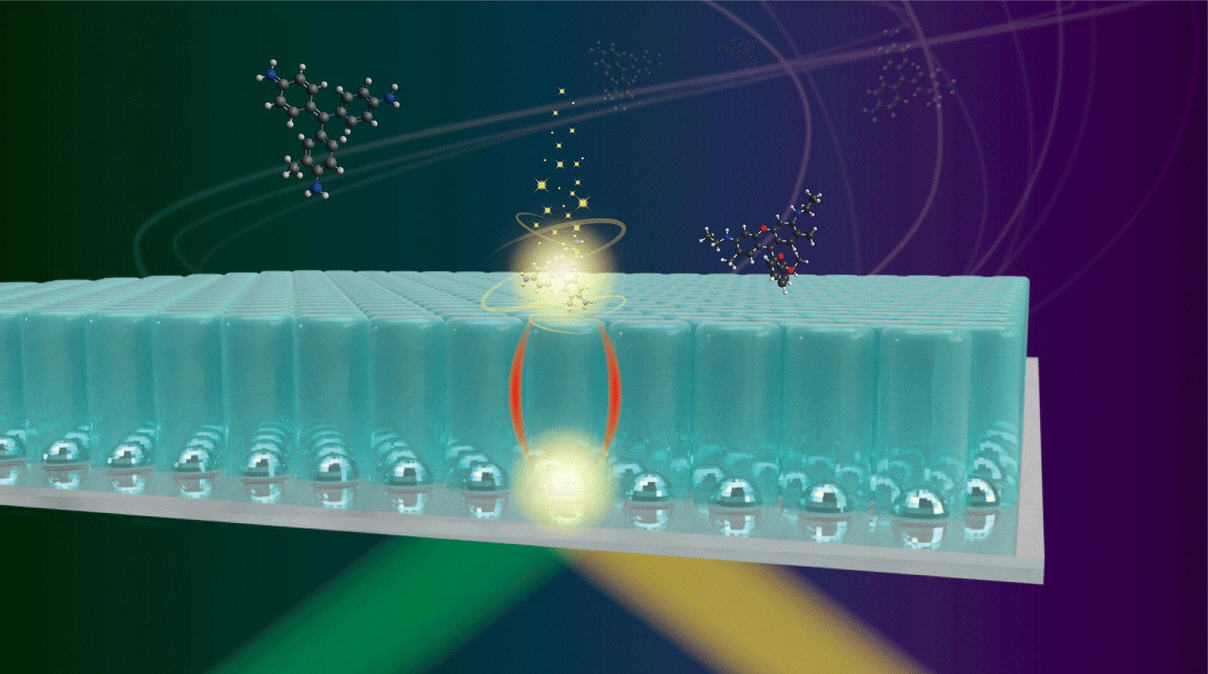
日本大阪大学(Osaka University, Osaka, Japan)的研究人员开发了一种新方法,利用密集随机排列的银纳米岛和保护性二氧化硅层来增强荧光和拉曼光谱信号。这一突破在不损坏细胞的情况下大大增强了检测能力,为环境监测和医疗诊断提供了潜在的应用。
先进光谱学技术(Advanced Spectroscopy Techniques)
今天的生物学家可以用比传统光学显微镜更先进的工具来探索活细胞内部复杂的结构。像荧光和拉曼光谱这样的技术已经成为无创监测生物过程的必要手段。这些方法使用光源,通常是激光来刺激荧光中的电子跃迁或拉曼光谱中的分子振动。
当前光谱方法的挑战(Challenges in Current Spectroscopic Methods)
尽管这些技术很有用,但它们也伴有挑战。荧光标签(Fluorescent tags)会干扰正常的细胞功能,而拉曼信号通常非常微弱。增加激光的功率或曝光时间来增强信号可能会损害敏感的生物分子。
为了克服这个问题,研究人员开发了这些方法的表面强化技术,使用金属衬底或纳米结构来放大信号。然而,这些强化也会给细胞完整性带来风险。
信号增强技术的突破(Breakthrough in Signal Enhancement)
现在,最近(2024年10月28日)发表在《光:科学与应用》(Light: Science & Applications)杂志上的一项研究——Takeo Minamikawa, Reiko Sakaguchi, Yoshinori Harada, Hiroki Tanioka, Sota Inoue, Hideharu Hase, Yasuo Mori, Tetsuro Takamatsu, Yu Yamasaki, Yukihiro Morimoto, Masahiro Kawasaki, Mitsuo Kawasaki. Long-range enhancement for fluorescence and Raman spectroscopy using Ag nanoislands protected with column-structured silica overlayer. Light: Science & Applications, 2024, 13(1): 299. DOI: 10.1038/s41377-024-01655-3. Pub Date 28 October 2024. 日本大阪大学等机构的科学家,描述了一种利用银纳米岛密集随机阵列远程增强荧光和拉曼信号的新方法。
参与此项研究的除了来自大阪大学的研究人员之外,还有来自日本德岛大学(Tokushima University, Tokushima, Japan)、日本京都立医科大学(Kyoto Prefectural University of Medicine, Kyoto, Japan)、日本科学技术振兴机构{PRESTO, Japan Science and Technology Agency (JST), Tokushima, Japan}、日本京都大学(Kyoto University, Kyoto, Japan)以及日本兵库县的Ushio公司技术工程部(Technology and Engineering Division, Ushio Inc., Hyogo, Japan)的研究人员。
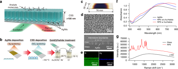
使用100 nm厚的柱状结构二氧化硅层将分析物分子与金属结构分开。这一层二氧化硅的厚度足以保护被研究的分子,但同时又足够薄,可以让金属层中的集体电磁振荡{称为等离子体激元(plasmons)}来增强光谱信号。
“我们证明了等离子体在金属中的影响范围可以超过100 nm,远远超出了传统理论的预测,”主要作者南川丈夫(Takeo Minamikawa)说。
对生物传感技术的启示(Implications for Biosensing Technology)
研究人员表明,使用这些生物相容的传感器基底可以将信号增加1000万倍。此外,由于金属纳米结构从不与所研究的分子直接接触,因此它们是可能被传统方法破坏的生物系统的理想选择。
资深作者川﨑三津夫(Mitsuo Kawasaki)说:“我们的基材的化学稳定性和机械稳健性使它们适用于广泛的应用,包括环境污染物检测或医疗诊断。”
此外,传感器衬底可以使用一种称为溅射(sputtering)的薄膜制造技术快速大规模地生产。因此,在工业和医疗保健环境中部署新的生物传感设备时,成本会更低。
本研究得到了日本学术振兴会{15K12519/MEXT | Japan Society for the Promotion of Science (JSPS)、19K22969/MEXT | Japan Society for the Promotion of Science (JSPS)、21H01847/MEXT | Japan Society for the Promotion of Science (JSPS)、22H00303/MEXT | Japan Society for the Promotion of Science (JSPS) }以及日本科学技术振兴机构{JPMJPR17PC/MEXT | JST | Precursory Research for Embryonic Science and Technology (PRESTO)}的资助。
上述介绍,仅供参考。欲了解更多信息,敬请注意浏览原文或者相关报道。
We demonstrate long-range enhancement of fluorescence and Raman scattering using a dense random array of Ag nanoislands (AgNIs) coated with column-structured silica (CSS) overlayer of over 100 nm thickness, namely, remote plasmonic-like enhancement (RPE). The CSS layer provides physical and chemical protection, reducing the impact between analyte molecules and metal nanostructures. RPE plates are fabricated with high productivity using sputtering and chemical immersion in gold(I)/halide solution. The RPE plate significantly enhances Raman scattering and fluorescence, even without proximity between analyte molecules and metal nanostructures. The maximum enhancement factors are 107-fold for Raman scattering and 102-fold for fluorescence. RPE is successfully applied to enhance fluorescence biosensing of intracellular signalling dynamics in HeLa cells and Raman histological imaging of oesophagus tissues. Our findings present an interesting deviation from the conventional near-field enhancement theory, as they cannot be readily explained within its framework. However, based on the phenomenological aspects we have demonstrated, the observed enhancement is likely associated with the remote resonant coupling between the localised surface plasmon of AgNIs and the molecular transition dipole of the analyte, facilitated through the CSS structure. Although further investigation is warranted to fully understand the underlying mechanisms, the RPE plate offers practical advantages, such as high productivity and biocompatibility, making it a valuable tool for biosensing and biomolecular analysis in chemistry, biology, and medicine. We anticipate that RPE will advance as a versatile analytical tool for enhanced biosensing using Raman and fluorescence analysis in various biological contexts.
https://blog.sciencenet.cn/blog-212210-1458063.html
上一篇:纳米塑料可以降低抗生素的有效性
下一篇:化学家打破了已有百年历史的规定,教科书改写势在必行

