博文
滴血测癌:科学家开发出从一滴血中快速、负担得起的癌症检测方法  精选
精选
||
滴血测癌:科学家开发出从一滴血中快速、负担得起的癌症检测方法
诸平
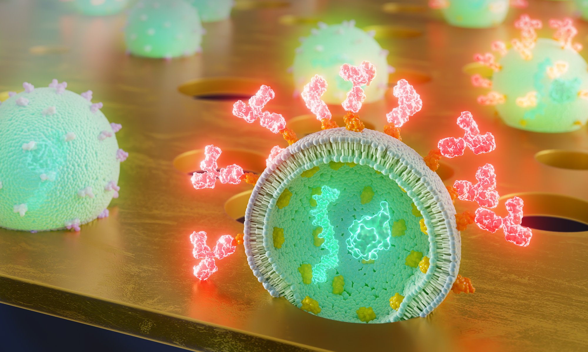
据美国罗彻斯特大学(University of Rochester, Rochester, NY, USA)2024年11月6日提供的消息,科学家开发出从一滴血中快速、负担得起的癌症检测方法(Scientists Develop Fast, Affordable Cancer Test From a Single Drop of Blood).该方法使用超薄膜来捕获被称为细胞外囊泡(extracellular vesicles简称EVs)的小包细胞物质。细胞释放数十亿小包细胞物质(EVs),进入血液、唾液和其他体液。这些细胞外囊泡(EVs)携带着关键的信息,包括来自其起源细胞的蛋白质和遗传物质,提供了对身体当前状态的洞察。
科学家们看到了EVs在诊断和治疗方面的巨大潜力,但在开发既快速又具有成本效益的方法方面面临诸多挑战。
2024年10月2日在《Small》杂志上发表的一项研究——Samuel N. Walker, Kilean Lucas, Marley J. Dewey, Stephen F. Badylak, George S. Hussey, Jonathan Flax, James L. McGrath. Rapid Assessment of Biomarkers on Single Extracellular Vesicles Using “Catch and Display” on Ultrathin Nanoporous Silicon Nitride Membranes. Small, 02 October 2024. DOI: 10.1002/smll.202405505. https://doi.org/10.1002/smll.202405505. 在此研究中,罗切斯特大学的研究人员与美国匹兹堡大学(University of Pittsburgh, Pittsburgh, PA, USA)以及罗切斯特大学医疗中心(University of Rochester Medical Center, Rochester, NY, USA)的研究人员合作,他们概述了一种使用超薄膜轻松识别EVs的新方法,以便进行快速液体活检。这种方法被称为液体活检的捕捉和显示(catch and display for liquid biopsy简称CAD-LB),有望快速、经济地诊断癌症,并评估用于治疗疾病的疗法的进展。
简化液体活检(Simplifying Liquid Biopsies)
生物医学工程教授、该研究的负责人詹姆斯·麦格拉斯(James McGrath)说:“通过在血液或其他体液样本中寻找这些细胞外囊泡及其携带的生物标志物,你可以找到身体出现问题的重要线索。这个想法已经存在了一段时间,但以前它需要许多纯化步骤才能将EVs与生物流体的其他成分分离开来。CAD-LB更简单和快速,这使其具有临床应用的潜力,这是更复杂的方法所缺乏的。”
该团队开发了超薄膜,其孔径正好可以捕捉EVs。一旦采集了血液样本,它就会被迅速处理,用移液管注射到膜上,然后在显微镜下直接分析。通过计算被评估疾病的生物标志物发光的小孔数量,使用者就可以快速估计疾病在体内的流行程度。
检测免疫蛋白和定制治疗方案(Detecting Immune Proteins and Tailoring Treatments)
上述论文除了概述CAD-LB方法外,该研究还证明了该方法识别EVs上关键免疫调节蛋白(immune modulatory proteins)的能力。这些蛋白质在帮助身体对抗肿瘤方面发挥着重要作用,并且可以预测患者对免疫疗法(immunotherapies)的反应。
罗切斯特大学医学中心泌尿外科(University of Rochester Medical Center’s Department of Urology)的研究助理教授、上述论文的合著者乔纳森·弗拉克斯(Jonathan Flax)说:“CAD-LB目前足够敏感,可以检测到一些处于可治愈阶段的癌症,这表明该技术具有癌症筛查的潜力。它也可以用来预测患者对免疫疗法的特异性选择,这种疗法指导免疫系统靶向并消除癌细胞。”
本研究得到了罗切斯特大学技术发展基金(University of Rochester Technology Development Fund)、罗彻斯特大学威尔莫特癌症研究所鲨鱼池试点项目(University of Rochester Wilmot Cancer Institute Shark Tank Pilot program)、美国国立卫生研究院(R01 EB031581/GF/NIH HHS/United States; TL1 TR001858/GF/NIH HHS/United States)以及美国国防部(CA170373/DOD)的资助或支持。
上述介绍,仅供参考。欲了解更多信息,敬请注意浏览原文或者相关报道。
New liquid biopsy method offers avenue to quick, affordable cancer diagnosis
Extracellular vesicles (EVs) are particles released from cells that facilitate intercellular communication and have tremendous diagnostic and therapeutic potential. Bulk assays lack the sensitivity to detect rare EV subsets relevant to disease, and while single EV analysis techniques remedy this, they are often undermined by complicated detection schemes and prohibitive instrumentation. To address these issues, a microfluidic technique for EV characterization called "catch and display for liquid biopsy (CAD-LB)" is proposed. In this method, minimally processed samples are pipette-injected and fluorescently labeled EVs are captured in the nanopores of an ultrathin membrane. This enables the rapid assessment of EV number and biomarker colocalization by light microscopy. Here, nanoparticles are used to define the accuracy and dynamic range for counting and colocalization. The same assessments are then made for purified EVs and for unpurified EVs in plasma. Biomarker detection is validated using CD9 and Western blot analysis to confirm that CAD-LB accurately reports relative protein expression levels. Using unprocessed conditioned media, CAD-LB captures the known increase in EV-associated ICAM-1 following endothelial cell cytokine stimulation. Finally, to demonstrate CAD-LB's clinical potential, EV biomarkers indicative of immunotherapy responsiveness are successfully detected in the plasma of bladder cancer patients treated with immune checkpoint blockade.
https://blog.sciencenet.cn/blog-212210-1458884.html
上一篇:康奈尔大学的突破可能意味着电池爆炸时代的终结
下一篇:睡眠对降低痴呆风险至关重要吗?
