博文
Development bioorthogonal SERS imaging probes
|
ChongguiQiu1【邱崇贵】ZiyiCheng1【程子译】Chuanzhu Lv*【吕传柱】Rui Wang*【王锐】Fabiao Yu*【于法标】
1 These authors contributed equally to this work.
The First Affiliated Hospital of Hainan Medical University, Key Laboratory of Emergency and Trauma, Ministry of Education, Key Laboratory of Hainan Trauma and Disaster Rescue, Institute of Functional Materials and Molecular Imaging, College of Pharmacy, College of Emergency and Trauma, Hainan Medical University, Haikou, 571199, China
Available online 9 March 2021.
https://doi.org/10.1016/j.cclet.2021.03.016
ABSTRACT
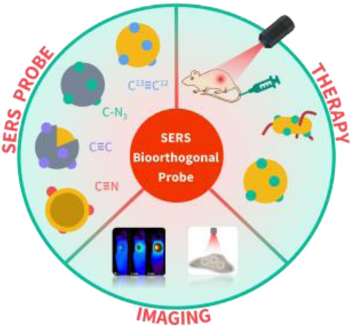
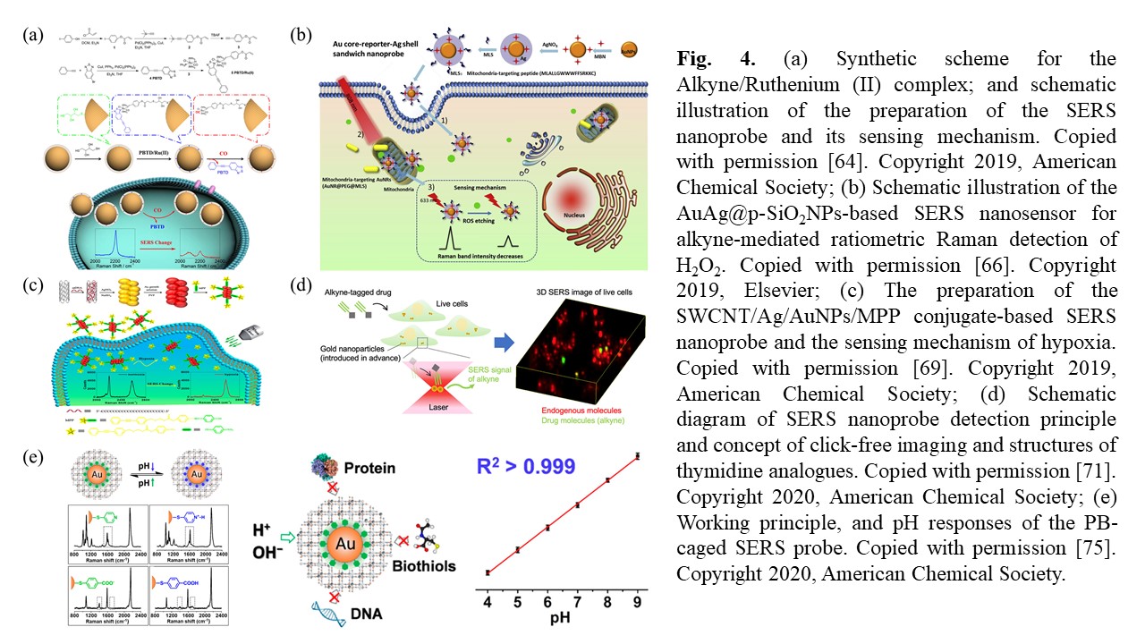
Living-cell imaging demands high specificity, sensitivity, and minimal background interference to the targets of interest. However, developing a desirable imaging probe that can possess all the above features is still challenging. The bioorthogonal surface-enhanced Raman scattering (SERS) imaging has been recently emerged through utilizing Raman reporters with characteristic peaks in Raman-silent region of cells (1800-2800 cm-1), which opens a revolutionary avenue for living-cell imaging with multiplexing capability. In this review, we focus on the recent advances in the technology development and the biological and biomedical applications of the living-cell bioorthogonal SERS imaging technique. After introduction of fundamental principles for bioorthogonal tag or label, we present applications for visualization of various intracellular components and environment including proteins, nucleic acids, lipids, pH and hypoxia, even for cancer diagnosis in tissue samples. Then, various bioorthogonal SERS imaging-guided therapy strategies have been discussed such as phototherapy and surgery. In conclusion, this strategy has great potential to be a flexible and robust tool for visualization detection and diseases diagnosis.
Graphical Abstract

Conclusion and perspective
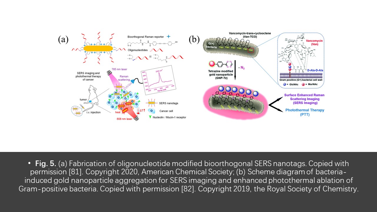
In the previous decade, many significant advances and technique development have been obtained in the field of bioorthogonal SERS imaging, which attracts much attention for living-cell imaging and whole-tissue studies. The most attractive feature is that the bioorthogonal Raman molecules can directly label the targets as well as ignore the impact on normal physiological activities, which makes the in situ and real-time visualization possible. In contrast, the common fluorophore or nanoparticle or enzyme are often too big in size to be metabolized, and usually interfere the normal physiological functions and processes, which limits their application in biological and biomedical research. Since SERS technique exhibits high sensitivity, multiple-detection capability, and resistance to photobleaching, the integration of SERS detection and bioorthogonal label will be an excellent strategy for visualization of living cells and tissues, even for the disease diagnosis.
It is worth noting that SERS stable labeling might alter the normal chemical and physiological properties of the substrate, even it has great potential in biomedicine, cell biology and environmental microbiology. Thus, the application of this strategy should be used with caution. Cell imaging can be expanded by integrating the types of bioorthogonal SERS tags into biomolecules through triple bond functional groups. This strategy not only has the labeling ability that does not interfere with the chemical properties of the substrate, but also has the properties of multiplexing imaging beyond traditional fluorescence imaging, which will make expected contribution for biological imaging. Although further improvement is desired for this strategy, the methodological development and biological and biomedical application have already demonstrated the potential in future research. We expect that bioorthogonal SERS imaging holds the prospect to monitor the specific cellular functions as the complement.
https://blog.sciencenet.cn/blog-2438823-1277640.html
上一篇:Probe for H2O2 Injuries in Pulmonary Fibrosis
下一篇:Fluorogenic Probe for Hepatocellular Carcer Chemotherapy
全部作者的其他最新博文
- • Triphenylamine-AIE Materials for Cancer Theranostics
- • Fluorescent Probe forRatiometric Monitoring of Peroxynitrite
- • Fluorescence Probe for Pathological Stages of Wound Healing
- • Macrophage M2 polarization to neurological damage
- • SERS-RCA biosensor for profiling dual miRNAs
- • a glutathione-activated near-infrared fluorescent probe