博文
Two-photon fluorescent probe for carboxylesterase 2 in mice
|
Visualization of carboxylesterase 2 with a near-infrared two-photon fluorescent probe and its potential evaluation of anticancer drug effects in orthotopic colon carcinoma mice model
Yan Wang【王艳】,ab Feifei Yu,b Xianzhu Luo【罗贤柱】,ab Mingshun Li【李明顺】,ab Linlu Zhao【赵琳璐】,*b and Fabiao Yu【于法标】*a
a. The Key Laboratory of Life-Organic Analysis, Key Laboratory of Pharmaceutical Intermediates and Analysis of Natural Medicine, College of Chemistry and Chemical Engineering, Qufu Normal University, Qufu 273165, China.
b. Institute of Functional Materials and Molecular Imaging, Key Laboratory of Emergency and Trauma, Ministry of Education, College of Pharmacy, Key Laboratory of Hainan Trauma and Disaster Rescue, College of Clinical Medicine, College of Emergency and Trauma, Hainan Medical University, Haikou 571199, China.
https://doi.org/10.1039/D0CC00297F Chemical Communications, 2020, 56, 4412 - 4415
We establish a near-infrared two-photon fluorescent probe for the detection of CE2 with high selectivity and sensitivity. This probe exhibits low cytotoxicity and superior tissue penetration ability for evaluating real-time activity of CE2 in living cells, in cancer tissues, and in colon carcinoma mice model.
Scheme 1 Illustration of the molecular structure DCM-CES2 and its proposed fluorescence response mechanism toward CE2.

Fig. 1 Spectral characterization of DCM-CES2 (10 μM). a) UV-Vis absorption spectrum with CE2 (0 - 10 μg/mL) for 10 min. b) The fluorescence spectrum with CE2 (0 - 15 μg/mL) for 10 min. c) Linear relationship between the fluorescence intensity and the activity of CE2. d) Time-dependent kinetic measurement of the fluorescent response to CE2 (5 μg/mL). e) Selectivity towards various species. Blank, Paraoxonase (PON1 and PON2, 10 μg/mL), Human serum albumin (HSA, 0.5 mg/mL), Bovine serum albumin (BSA, 0.5 mg/mL), Acetylcholinesterase (AChE, 0.1 μg/L), Butyrylcholinesterase (BChE, 20 U/L), CES1 (10 μg/mL), Loperamide (LAP, 100 μg/mL), Bis(4-nitrophenyl) phosphate (BNPP, 100 μg/mL), and CE2 (10 μg/mL). f) Lineweaver-Burk plot for the enzymatic reaction. The Michaelis-Menten equation was expressed as: V = Vmax [probe]/(Km + [probe]), where V was the reaction rate, [probe] referred to the probe concentration (substrate), and Km was the Michaelis constant. λex = 560 nm.
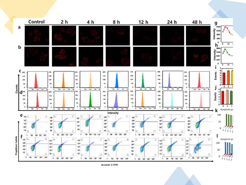
Fig. 2 Two-photon confocal fluorescence imaging (λex = 830 nm, λem = 650 - 750 nm) and flow cytometry assay (λex = 560 nm, λem = 650 - 750 nm) for detection of CE2 in HT-29 cells. a - b) HT-29 cells pretreated with CPT-11 and CAPE, respectively, and then incubated with DCM-CES2. c - d) Corresponding flow-cytometric analysis to a and b, respectively. e - f) PE Annexin V/7-AAD assays of HT-29 cells in a and b, respectively, using flow cytometry, Q1: necrosis cells, Q2: late apoptotic cells, Q3: early apoptotic cells, Q4: survival cells. g - h) Mean fluorescence intensities of the images in a and b, respectively. i - j) Mean values of c and d, respectively. k - l) Apoptosis percentages of e and f, respectively. The data were shown as mean (± s.d.).
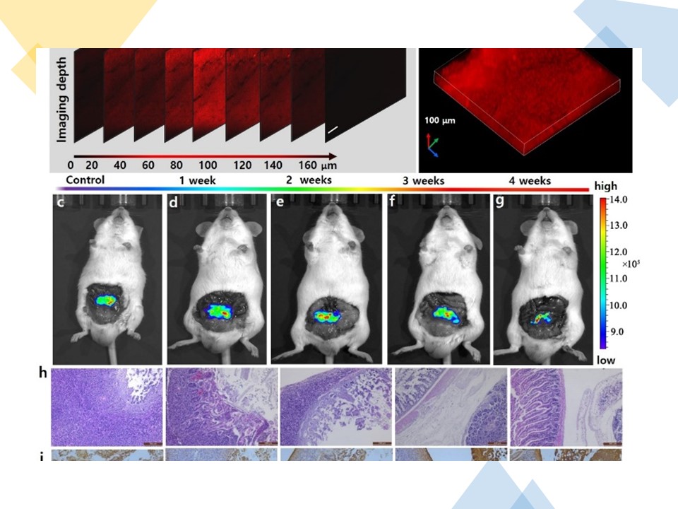
Fig. 3 a) The confocal z-scan TP imaging at different depths. b) 3D fluorescence image in a. scale bar: 100 μm. c - g) Evaluation of CE2 in orthotopic tumor-bearing mice model treated with CPT-11 by tail intravenous injection (20 mg/kg) for 0 - 4 weeks and fluorescence images of CE2 with DCM-CES2. h) H&E stained colonic cancer tissues histopathology images. i) Regional TUNEL staining in the colonic cancer tissues. k) Masson's stained slice of colonic cancer tissues.
In conclusion, we have successfully designed and synthesized a NIR-emissive two-photon fluorescent probe DCM-CES2 for endogenous CE2 detection in living cells and in vivo. The probe offers a switch-on behavior for the detection of CE2 activity under simulated physiological systems with a large Stokes shift of 125 nm and a low LOD as 0.087 μg/mL. Cell experiments illustrate that DCM-CES2 can realize specific detection of endogenous CE2 with low cytotoxicity and satisfactory cell membrane permeability. Tissue fluorescence imaging exhibits high resolution at penetration depth up to 160 μm due to its excellent NIR-emissive TP properties. DCM-CES2 has been applied for real-time monitoring of CE2 activity during the activation process of anticancer prodrugs CPT-11 and CAPE in cells. The probe also achieves in situ and in vivo evaluation of therapeutic efficacy of anticancer prodrug CPT-11 at tumour lesion in orthotopic colonic mice model. The results of DCM-CES2 reveal that this NIR two-photon probe holds potential advantages for the further study of the biological function of CE2 in complex systems.
https://blog.sciencenet.cn/blog-2438823-1229709.html
上一篇:海南医学院2020年临床医学学术型硕士和博士研究生招生
下一篇:Fluorescent Probes for HClO in Osteoarthritis Mouse Model
全部作者的其他最新博文
- • Triphenylamine-AIE Materials for Cancer Theranostics
- • Fluorescent Probe forRatiometric Monitoring of Peroxynitrite
- • Fluorescence Probe for Pathological Stages of Wound Healing
- • Macrophage M2 polarization to neurological damage
- • SERS-RCA biosensor for profiling dual miRNAs
- • a glutathione-activated near-infrared fluorescent probe