博文
Self-assembled nanomaterials for phototherapy of cancer
|
Self-assembled nanomaterials for enhanced phototherapy of cancer
Linlu Zhao【赵琳璐】, Yanlong Xing【邢艳珑】, Rui Wang【王锐】, FeiFei Yu【于飞飞】,* Fabiao Yu【于法标】*
Institute of Functional Materials and Molecular Imaging, Key Laboratory of Emergency and Trauma, Ministry of Education, College of Pharmacy, Key Laboratory of Hainan Trauma and Disaster Rescue, College of Clinical Medicine, College of Emergency and Trauma, Hainan Medical University, Haikou 571199, China
ACS Appl. Bio Mater.
Publication Date:November 28, 2019
https://doi.org/10.1021/acsabm.9b00843
ABSTRACT: Following nature’s inspiration to assemble functional building blocks into exquisite nanostructures is emerging as a fascinating research field and has been widely applied in biomedical sciences. Owing to their excellent features of structural and functional diversity, high biocompatibility and dynamic adjustability, the self-assembled nanoarchitectures come to play a pivotal role in fighting against cancer. This review outlines the most up-to-date developments in constructing phototherapeutic nanomaterials for photodynamic and photothermal therapy (PDT and PTT) of tumors, with emphasis on design ideas, building blocks, and advantageous characteristics of self-assembly. The prominent antitumor activities achieved by these phototherapeutic nanomaterials are also discussed in-depth, along with the connections between the specific nanostructures and their unique properties, providing a comprehensive understanding of the self-assembled nanomaterials in improving the outcomes of PDT and PTT. The goal of this review is to highlight the significance of self-assembled nanomaterials in enhancing phototherapeutic efficacy and to promote its growth in a diverse variety of research fields ranging from nanoscience and material science to biomedicine and clinical medicine.
KEYWORDS: Supramolecular self-assembly, functional nanomaterials, photodynamic therapy, photothermal therapy, cancer theranostics.

Figure 1. Schematic illustration showing self-assembly of functional building blocks in the construction of phototherapeutic nanomaterials through non-covalent interactions for PDT, PTT and the diagnostic modalities such as PAI and MRI for imaging-guided phototherapy of cancer.
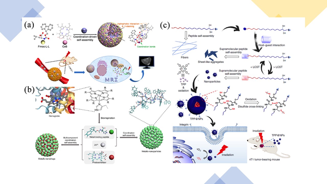
Figure 2. (a) Schematic illustration of the fabrication process of Fmoc-LL/Mn2+ nanoparticles (FMCNPs) via coordination-driven self-assembly and their responsive disassembly for MRI-guided PDT. Reproduced with permission from ref. 59. Copyright 2018 American Chemical Society. (b) Construction of metallo-nanodrugs through cooperative coordination of small peptides and photosensitizers in the presence of zinc ions. Reproduced with permission from ref. 61. Copyright 2018 American Chemical Society. (c) Schematic illustrations of the programmable peptide self-assembly and PDT process. Solid arrows indicate experimental procedure and hollow arrows refer to self-assemblies or structural details. Reproduced with permission from ref. 62. Copyright 2019 Nature Publishing Group.
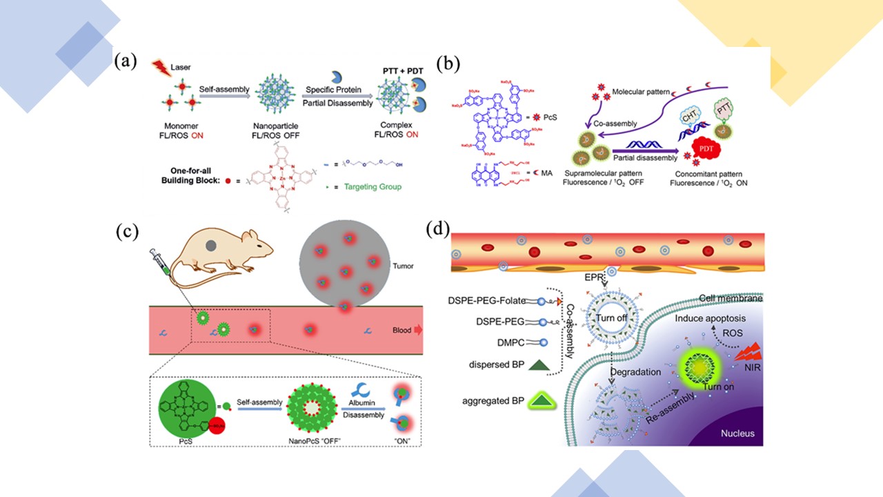
Figure 3. (a) Schematic illustration of the dynamic Pc assembly and partial disassembly processes as well as the switchable photoactivity. Reproduced with permission from ref. 66. Copyright 2018 American Chemical Society. (b) Schematic illustration of the construction of a nanotheranostic agent based on supramolecular interaction between PcS and MA and its nucleic-acid-driven activatable properties for fluorescent imaging and PDT synergized with PTT and CHT. Reproduced with permission from ref. 67. Copyright 2018 American Chemical Society. (c) Schematic illustration of the dynamic PcS assembly and disassembly processes as well as the switchable photoactivities for in vivo fluorescence imaging and tumor-targeted PDT. Reproduced with permission from ref. 68. Copyright 2019 American Chemical Society. (d) Illustration of coassembled AIE-PS@liposomes composed of AIE-PS and lipid for antitumor in PDT. Reproduced with permission from ref. 69. Copyright 2019 American Chemical Society.
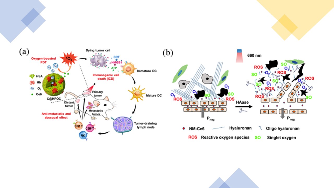
Figure 4. (a) Schematic depiction of oxygen-augmented immunogenic PDT utilizing a bioinspired hybrid protein oxygen nanocarrier with Ce6 loaded (C@HPOC) for eliciting the anti-metastatic and abscopal effect. Reproduced with permission from ref. 81. Copyright 2018 American Chemical Society. (b) Schemes showing the effects of HAase on the modulation of tumor microenvironment, resulting in enhanced efficacy of in vivo PDT cancer treatment via improving tumor oxygenation and promoting EPR effect for NPs. Reproduced with permission from ref. 82. Copyright 2016 American Chemical Society.
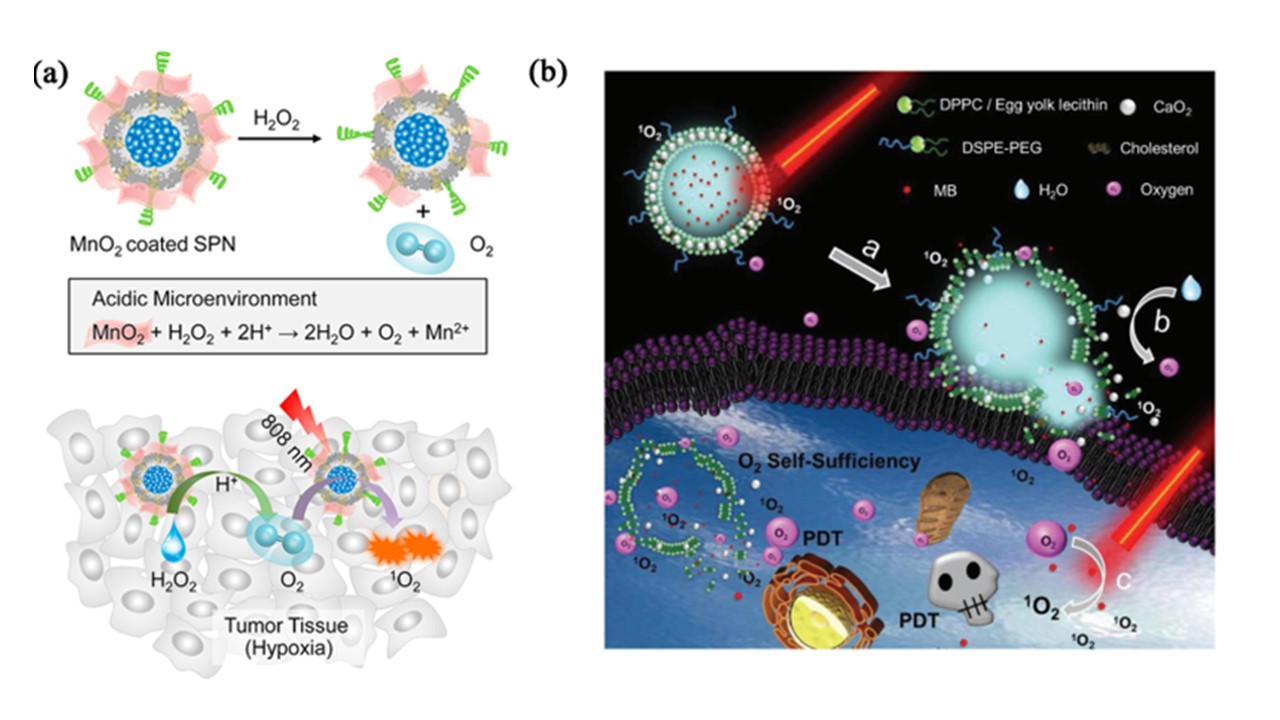
Figure 5. (a) Schematic illustration for the H2O2-responsive mechanisms of SPN-M1 and the detailed mechanism of SPN-M1 for amplified photodynamic therapy in tumor. Reproduced with permission from ref. 84. Copyright 2018 American Chemical Society. (b) Structure illustration of LipoMB/CaO2 nanoplatform for O2 self-sufficient PDT. Reproduced with permission from ref. 85. Copyright 2017 John Wiley and Sons.
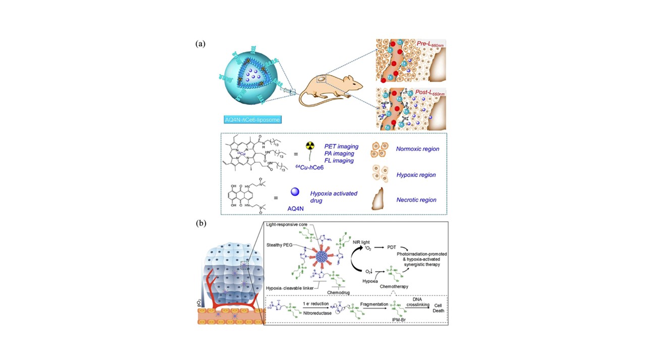
Figure 6. (a) A scheme illustrating the chemical composition of AQ4N-hCe6-liposome, which can serve as a multifunctional theranostic agent for multimodal imaging and PDT-induced, hypoxia-activated cancer therapy. Reproduced with permission from ref. 89. Copyright 2017 American Chemical Society. (b) Schematic of SPNpd for hypoxia-activated synergistic PDT and chemotherapy. Reproduced with permission from ref. 88. Copyright 2019 John Wiley and Sons.
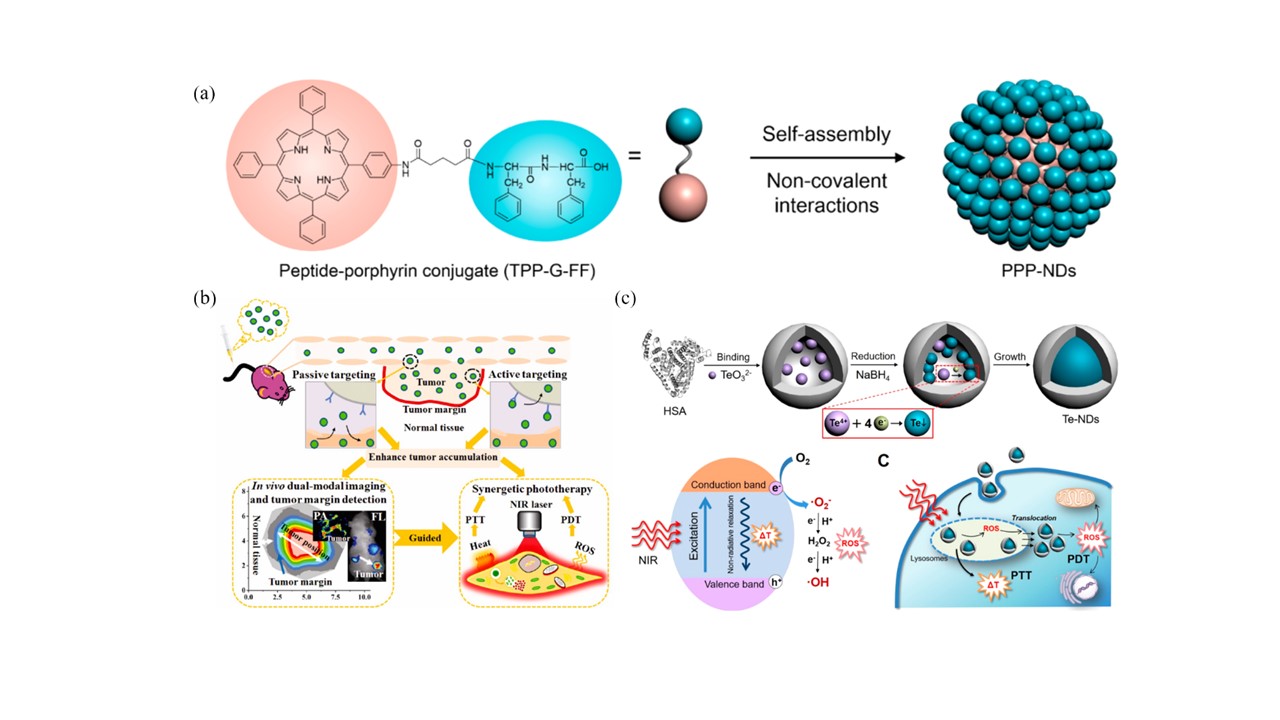
Figure 7. (a) Self-assembly of a peptide-porphyrin conjugate (TPP-G-FF) into photothermal peptide-porphyrin nanodots (PPP-NDs). Reproduced with permission from ref. 105. Copyright 2017 American Chemical Society. (b) Schematic illustration of HSA-ICG NPs for in vivo dual-modal imaging, tumor margin detection, and simultaneous PDT/PTT treatments. Reproduced with permission from ref. 106. Copyright 2014 American Chemical Society. (c) Schematic illustration of Te-NDs preparation through albumin nanoreactor approach, photoconversion mechanism for Te-NDs to generate simultaneous photothermal effect and ROS under single wavelength NIR light irradiation, and their intracellular synergistic photothermal and photodynamic treatments. Reproduced with permission from ref. 107. Copyright 2017 American Chemical Society.
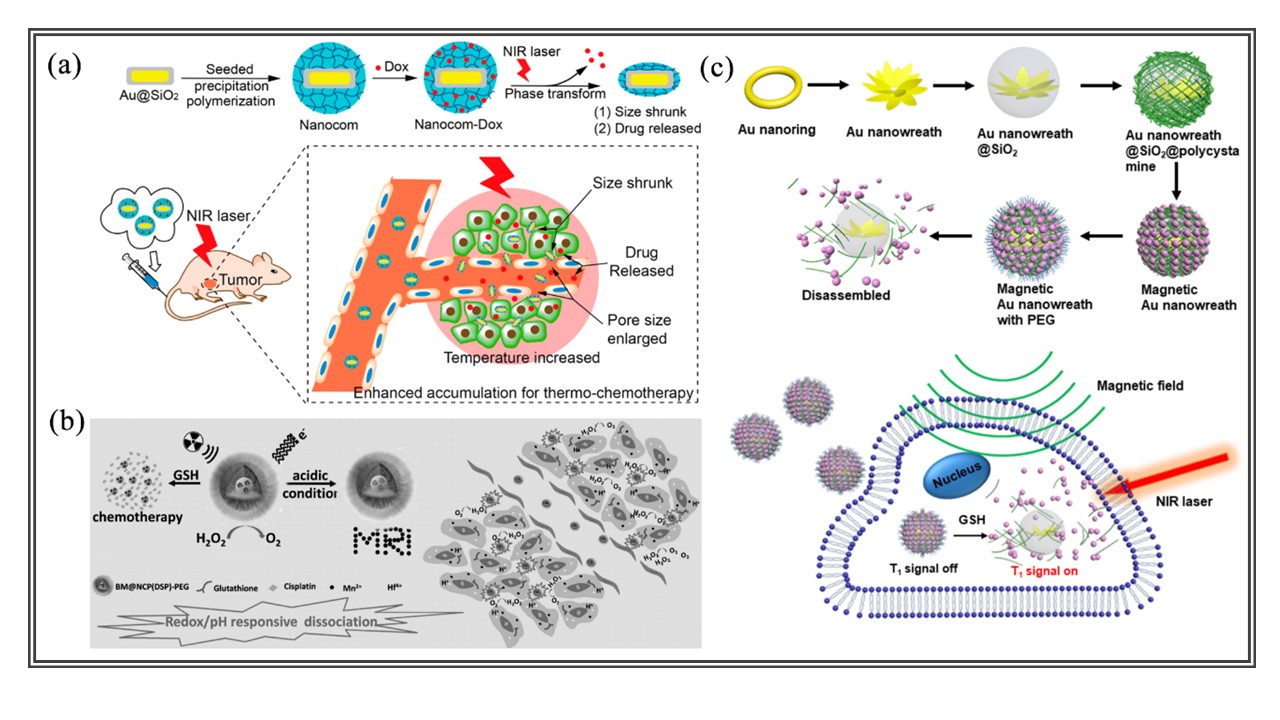
Figure 8. (a) Nanocomposite formulation process and NIR laser induced targeted thermo-chemotherapy using the Nanocomposite. Reproduced with permission from ref. 111. Copyright 2014 American Chemical Society. (b) A scheme illustrating the redox/pH responsive behaviors of BM@NCP(DSP)-PEG composite NPs in the tumor microenvironment. Such NPs are responsive to reduced pH, reductive environment, and existence of tumor endogenous H2O2, enabling tumor-specific imaging, drug release, hypoxia relief, and enhanced cancer chemoradiotherapy. Reproduced with permission from ref. 112. Copyright 2017 John Wiley and Sons. (c) Schematic illustration of the synthesis of magnetic gold nanowreath and their applications as GSH-responsive T1-weighted imaging contrast agents and photothermal agents. Reproduced with permission from ref. 113. Copyright 2018 American Chemical Society.
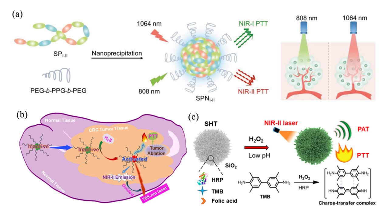
Figure 9. (a) Design and synthesis of SPNI-II: Synthesis of SPNI-II via nanoprecipitation; Scheme that illustrates the deeper tissue penetration of 1064 nm relative to that of 808 nm. Power density: 808 nm, 0.3 W/cm2; 1064 nm, 1 W/cm2. Reproduced with permission from ref. 121. Copyright 2018 John Wiley and Sons. (b) Schematic illustration of Nano-PT as an activatable photothermal agent for NIR-II fluorescence-guided therapy of H2S-rich colorectal cancers. Reproduced with permission from ref. 122. Copyright 2018 American Chemical Society. (c) Illustration of SHT formation and its H2O2 activation and acid enhancement for tumor-specific NIR-II photonanotheranostics. Reproduced with permission from ref. 123. Copyright 2019 American Chemical Society.

Figure 10. (a) Schematic illustration of the single-step sonication to synthesize DINPs. Photograph of mixture containing Dox, ICG, lecithin, and DSPE-PEG was before (left) and after (right) sonication. Reproduced with permission from ref. 127. Copyright 2013 American Chemical Society. (b) The mechanism of anti-tumor immune responses induced by PLGA-ICG-R837-based PTT in combination with checkpoint-blockade. Reproduced with permission from ref. 128. Copyright 2016 Nature Publishing Group. (c) Photosensitizer (Ce6)-loaded plasmonic gold vesicles (GVs) for trimodality fluorescence/thermal/photoacoustic imaging guided synergistic photothermal/photodynamic cancer therapy. (d) In vivo NIR fluorescence image of MDA-MB-435 tumor-bearing mice at pre-injection and post-injection of GV-Ce6; Thermal images of tumor-bearing mice exposed to 671 nm laser (2.0 W/cm2) for 6 min at post-injection of GV-Ce6. Red circles indicate the location of tumors. Reproduced with permission from ref. 129. Copyright 2013 American Chemical Society.
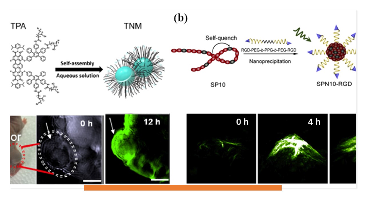
Figure 11. (a) Self-assemble mechanism of the TNMs and in vivo 3D PA imaging of tumor tissue at the highest signal intensity timepoint (12h). Reproduced with permission from ref. 141. Copyright 2017 American Chemical Society. (b) Synthesis of the targeted SPN (SPN10-RGD) and PA images of tumor after systemic administration of SPN10-RGD for 0, 4 and 24 h. Reproduced with permission from ref. 142. Copyright 2017 Elsevier B.V.
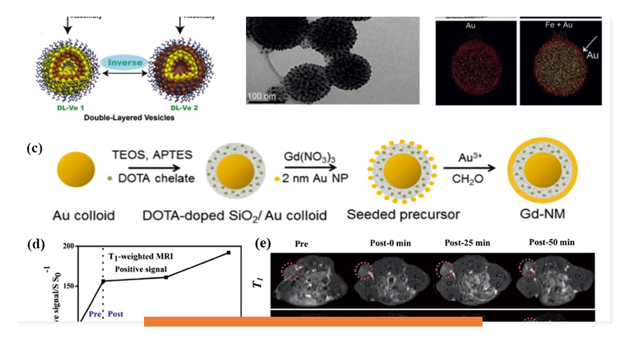
Figure 12. (a) Schematic illustration of the formation of the hierarchical self-assembly of the resulting three kinds of Janus amphiphilic NPs into double-layered plasmonic–magnetic vesicle 1. (b) TEM, SEM images, and TEM-element mapping images of the double-layered vesicle 1. Reproduced with permission from ref. 149. Copyright 2017 John Wiley and Sons. (c) Schematic representation of the MRI-active NM synthesis showing the stepwise synthesis process: the 50-nm-diameter gold colloids are coated with SCN-DOTA chelates embedded in a SiO2 shell, and then incubated in Gd(NO3)3 and 2 nm Au NP, followed by the growth of a continuous Au shell. Reproduced with permission from ref. 145. Copyright 2017 The American Association for the Advancement of Science. (d) Intensity change of T1-weighted and T2-weighted MR signal of FeS2-PEG at different times. (e) Corresponding MR images of mice, verifying the TME-responsive self-enhanced MRI behavior in vivo (the red dotted circles). Reproduced with permission from ref. 154. Copyright 2017 John Wiley and Sons.
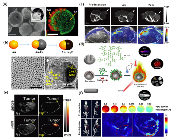
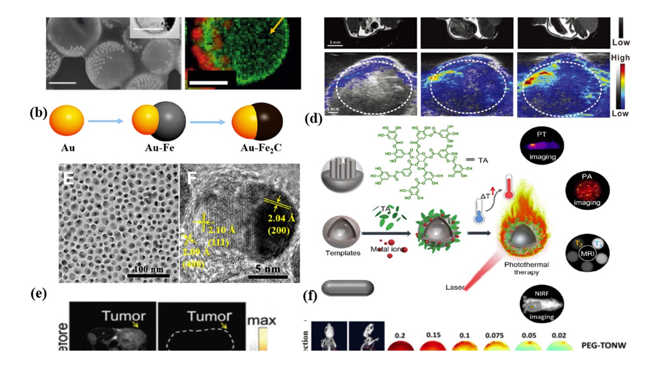
Figure 13. (a) SEM and TEM (inset) images of magneto-plasmonic JVs with spherical shapes. Reproduced with permission from ref. 158. Copyright 2016 John Wiley and Sons. (b) Schematic depiction of the synthetic process of Au−Fe2C JNPs; TEM (left) and HRTEM (right) images of Au−Fe2C JNPs. Reproduced with permission from ref. 159. Copyright 2017 American Chemical Society. (c) In vivo multimodal imaging of MR images and PA images of U87MG-tumor-bearing mice pre- and post- intravenous injection of PGM NPs. Reproduced with permission from ref. 160. Copyright 2017 John Wiley and Sons. (d) Schematic illustration of the cooperation of adhesive MITAs with diverse templates for advanced applications, including PTI, PAI, T1- and T2-weighted MRI, and near-infrared fluorescence (NIRF) imaging together with PTT. Reproduced with permission from ref. 161. Copyright 2018 American Chemical Society. (e) In vivo 1H- and 19F-MRI of the tumor-bearing mice before and after the injection of NCs. Reproduced with permission from ref. 162. Copyright 2018 American Chemical Society. (f) Left: CT images of a mouse before and after intravenous injection of PEG-TeO2/(NH4)xWO3 nanoribbons (PEG-TONW NRs) (20 mg kg−1); right: PA imaging of PEG-TONW NRs buffer solution and in vivo PA imaging of a tumor-bearing mouse before (up) and after (down) intravenous injection of PEG-TONW NRs. Reproduced with permission from ref. 164. Copyright 2019 American Chemical Society.
https://blog.sciencenet.cn/blog-2438823-1212020.html
上一篇:Glutathione fluorescent probe and surgery-guide in cancer
下一篇:selenocysteine fluorescent probe for thyroid diseases
全部作者的其他最新博文
- • Triphenylamine-AIE Materials for Cancer Theranostics
- • Fluorescent Probe forRatiometric Monitoring of Peroxynitrite
- • Fluorescence Probe for Pathological Stages of Wound Healing
- • Macrophage M2 polarization to neurological damage
- • SERS-RCA biosensor for profiling dual miRNAs
- • a glutathione-activated near-infrared fluorescent probe